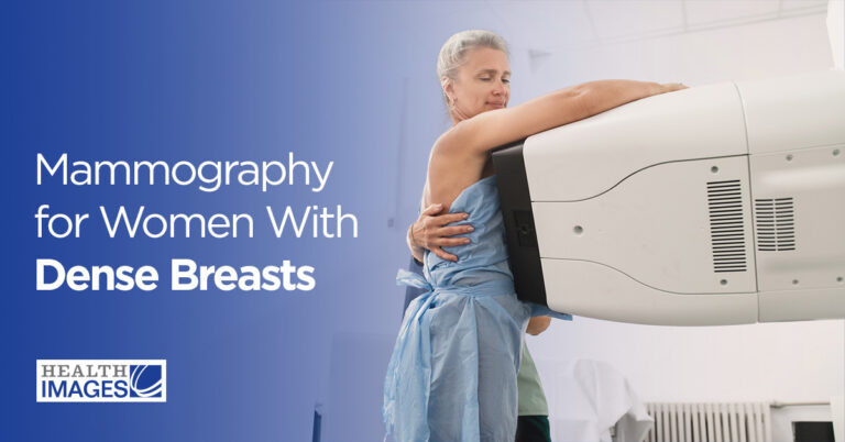
If you have dense breasts, supplemental breast cancer screening is an excellent way to gain peace of mind and be proactive about your health. Supplemental screening methods, such as an automated breast ultrasound (ABUS), can reveal abnormalities that dense tissue may hide on a standard mammogram. While annual mammograms remain the most important breast cancer screening tool, supplemental screening can increase your chances of early detection.
What Does It Mean to Have Dense Breasts?
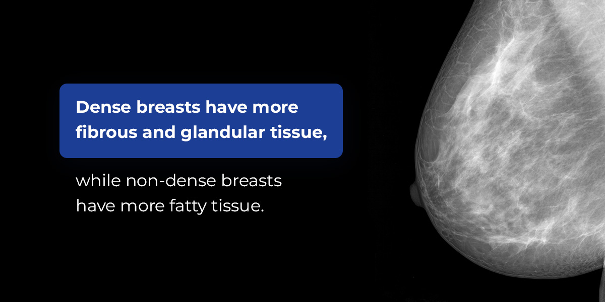
Breast density refers to the composition of the breast, or how much fatty tissue and connective tissue are present. Dense breasts have more glandular and fibrous tissue, and non-dense breasts are made up of more fatty tissue. Fibrous tissue provides structural support, and glandular tissue consists of milk glands and the ducts that transport milk following a pregnancy. These tissues are what give breasts their particular shape and firmness, and their relative amount varies by person.
Dense and non-dense breasts are completely normal, but they can impact how tissue and cancer cells appear on screens.
How to Tell if You Have Dense Breasts
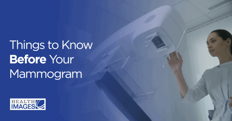
You can determine if you have dense breasts or primarily fatty breast tissue through a medical screening such as a mammogram, breast ultrasound or magnetic resonance imaging (MRI). Most women with dense breasts learn about their breast density following a mammogram.
Various Levels of Breast Density
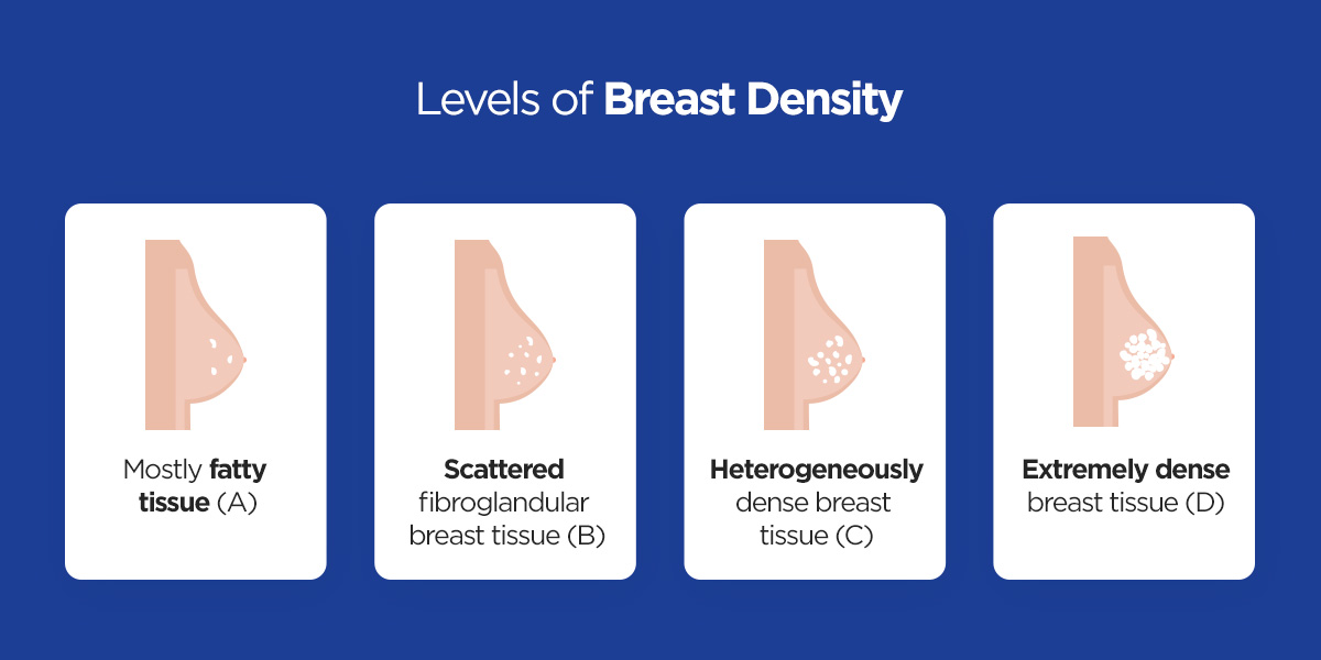
The Food and Drug Administration (FDA) requires mammography providers to inform each patient whether their breasts are “dense” or “not dense” in their mammogram reports. For a more detailed explanation of your breast density, you can ask your provider for your full mammography report. While simplified reports categorize density as dense or not dense, a detailed report reveals which of the following breast imaging reporting and data system (BI-RADS) categories your breasts are part of:
Mostly fatty tissue (A): If your breasts are categorized as mostly fatty tissue, this means that almost all of your breast tissue is fatty tissue.
Scattered fibroglandular breast tissue (B): Scattered fibroglandular breast tissue is a mix of fatty and dense tissue, but it’s mostly fatty.
Heterogeneously dense breast tissue (C): Heterogeneously dense breast tissue is also a mix of tissue, but it’s mostly dense tissue.
Extremely dense breast tissue (D): If you have extremely dense breast tissue, this indicates that almost all of your breast tissue is dense.
Approximately 80% of the population has a mix of scattered fibroglandular breast tissue or heterogeneously dense breast tissue, and 20% of the population has either mostly fatty or mostly dense tissue. Scattered fibroglandular breast tissue, heterogeneously dense breast tissue and extremely dense breast tissue can all impact mammogram results, making it important to seek additional screening options.
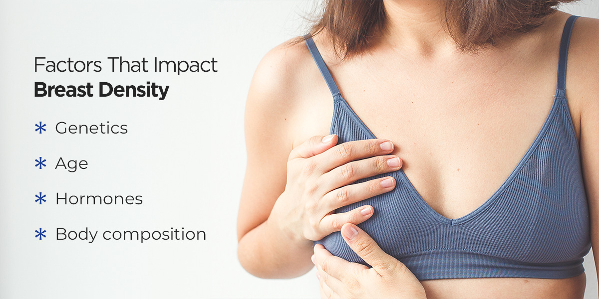
What Causes Higher Breast Density?
The following factors can impact breast density:
Genetics: A family history of breast density can increase your chances of having dense breasts.
Age: Density can decrease with age, but this can vary by person.
Hormones: Higher levels of estrogen and progesterone can increase breast density, while higher levels of testosterone can decrease density. Some women’s breast density increases with hormone replacement therapy (HRT).
Body composition: Higher breast density is often connected to a low body mass index.
How Breast Density Affects Cancer Detection
Breast density impacts cancer detection because it shows up as the same color on standard 2D mammograms. When a provider completes a mammogram, fatty tissue appears as black or dark gray, and connective and glandular tissue appears as white. Abnormalities such as tumors and benign conditions also appear white.
Dense breast tissue can make it harder for radiologists to distinguish between the tissue and abnormalities, essentially “hiding” or “masking” potential tumors.
Do Dense Breasts Increase the Chance of Cancer?
Having primarily dense breast tissue can slightly increase the risk of developing breast cancer because breast cancer cells can originate in glandular tissue, but the primary concern is how it impacts screening. Relying solely on standard 2D mammograms to detect cancer in dense tissue can increase the risk of false negatives. Supplemental screening methods help providers detect cancer earlier, giving you peace of mind and increasing positive treatment outcomes.
Advanced Mammography for Women With Dense Breasts
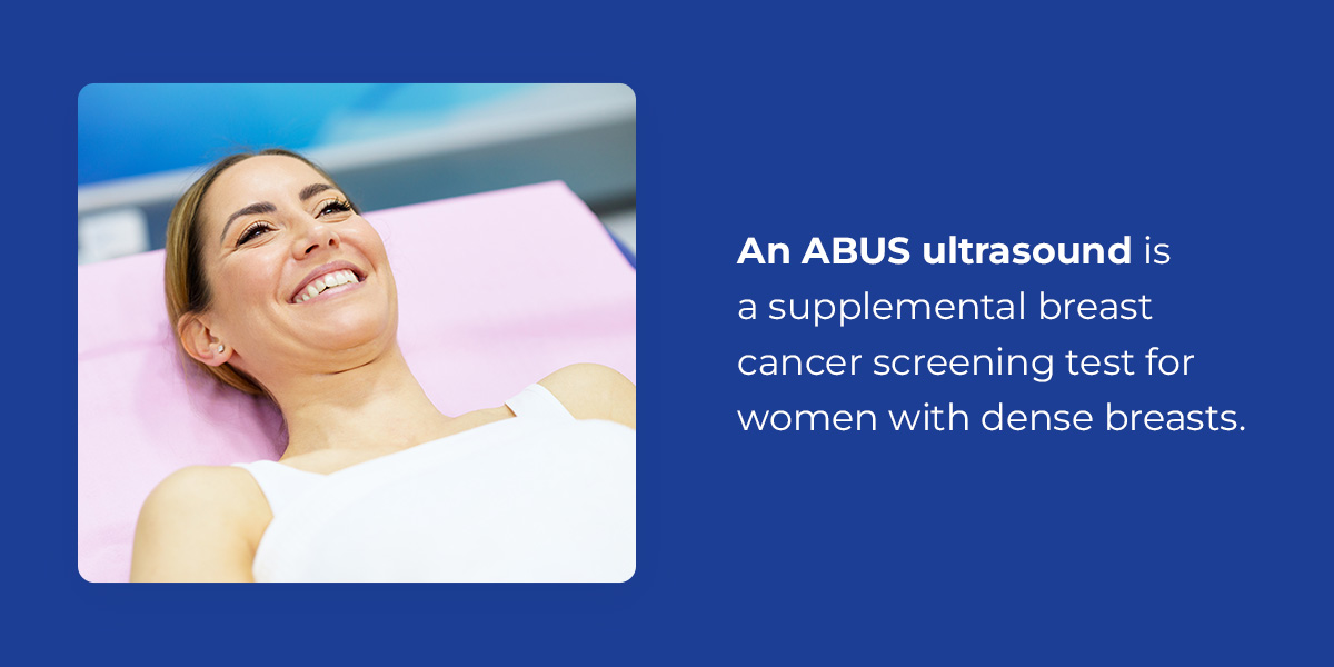
Supplemental screening options are available for women with dense breasts, such as an MRI, handheld ultrasound or an automated breast ultrasound. An ABUS ultrasound is a supplemental breast cancer screening test for women with dense breasts. It creates a 3D image of the breast with ultrasound technology, catching abnormalities that may not appear on a mammogram. This technology uses an automated scanner and sound waves that bounce off different tissues. The scanner converts the echoes into electronic signals, and a computer processes these signals into detailed 3D images.
Why Is an ABUS the Best Option for Screening Dense Breasts?
An ABUS is an excellent supplemental cancer screening method for dense breasts because it offers the following advantages:
Accuracy and clarity for dense breasts: An ABUS delivers more accurate results for dense breasts than a standard mammogram alone because cancer shows up as a darker or black color against dense white tissue. It can detect cancers that dense tissue might hide on a mammogram.
Standardized imaging: Since the ABUS test is automated, it reduces operator skill variations and inconsistencies between scans. This results in reproducible, standardized images your provider can easily review and compare to past or future exams.
Comprehensive evaluation: An ABUS also enables a more reliable, comprehensive evaluation of breast tissue. Its multi-layered view can help your provider detect cancer early, preventing unnecessary biopsies.
Comfort: ABUS screening does not require breast compression like mammograms do, making it a quicker, more comfortable experience.
No radiation: ABUS technology relies on sound waves instead of x-rays, making it a safer option if avoiding radiation exposure is important to you.
Who Are Good Candidates for ABUS Screening?
A standard mammogram is adequate for some women, while others require supplemental screening. ABUS is recommended for women who:
Have heterogeneously dense breast tissue or extremely dense breast tissue.
Are at a higher risk of breast cancer due to genetic factors or family history.
Have a high risk of breast cancer but cannot undergo MRI screening.
It’s important to remember that ABUS is an addition to mammograms, not a replacement. Some cancers, such as those involving microcalcifications, are only visible on standard mammograms. You may also need additional imaging for the underarm lymph node region because ABUS technology does not cover this area.
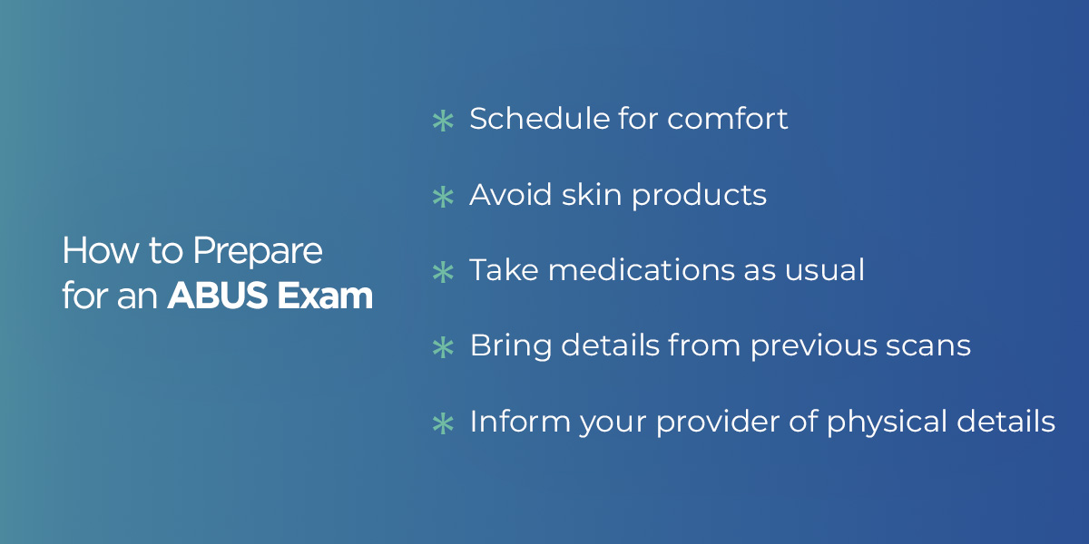
How to Prepare for an ABUS Exam
Use the following tips to prepare for an ABUS exam:
Schedule for comfort: If you experience breast tenderness or soreness around your period, try to schedule for the week before or after when tenderness is lower. This helps ensure your comfort during the exam.
Avoid skin products: Avoid applying oils, lotions or powders on your chest and breasts the day of your exam. These can interfere with ultrasound contact.
Take medications as usual: You can take your usual medications before your exam. If you are sensitive to pressure, you may take an over-the-counter pain reliever an hour before the screening.
Bring details from previous scans: List your previous breast imaging dates and locations, or bring outside images on a disc or via portal access. You may need to contact past imaging facilities to send your results to you or your ABUS provider. This enables the radiologist to compare past images to your current results.
Inform your provider of physical details: Let your provider know if you have limited mobility, breast implants or open wounds or skin irritation on your chest. You should also let them know if you have had a recent biopsy or surgery, or if you’re pregnant. ABUS is safe during pregnancy, but notifying the care team is helpful.
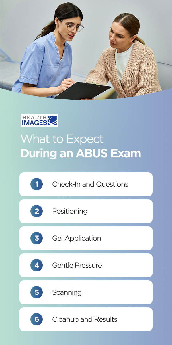
What to Expect During an ABUS Exam
An ABUS screening is noninvasive, taking about 15-20 minutes and involving the following steps:
1. Check-In and Questions
After checking in, you will change into a hospital gown and your technologist will explain the process and answer any questions you may have.
2. Positioning
The technologist will ask you to lie on your back on the exam table with your arm placed above your head, which places the breast comfortably on the side being scanned.
3. Gel Application
After helping you position on the table, the technologist will apply a gel or gel lotion to your breast to reduce friction and help ultrasound waves travel efficiently between the ultrasound probe and the body. Air gaps between the skin and the probe can absorb or reflect ultrasound waves, but the gel fills these gaps for optimal image quality. The gel may feel cool when first applied.
4. Gentle Pressure
The technologist will apply a large curved probe on your breast with gentle but firm pressure to ensure steady contact and an even scan. This pressure is different from mammogram compression, and most people find it more comfortable.
5. Scanning
The ABUS scan takes only a few minutes for each breast, automatically sweeping across sections of your breast. There are typically three to five passes per breast so the probe can catch images of the outer, center and inner portions. Your technologist will ask you to lie still and breathe normally during the scanning process.
You will remain covered as much as possible during your ABUS exam, and you can tell your technologist if you feel any discomfort during the process or need to pause. They can adjust pressure and positioning if needed.
6. Cleanup and Results
After the exam, you can wipe the gel off and resume normal activities right away. A radiologist will review your images after your visit, and you should receive results within a few days.
Your provider may schedule you for additional imaging if your results show any areas of concern, such as a cyst, mass or unusual calcification patterns. A handheld ultrasound can provide more detailed imaging if your provider needs to further evaluate abnormalities and determine if they are benign or require further action.
MRI Screening for Breast Cancer
In some cases, your provider may recommend an MRI instead of an ABUS. An MRI is a helpful supplemental screening option if you have additional risk factors, such as:
The BRCA1 or BRCA2 gene mutation
A first-degree relative who carries the BRCA1 or BRCA2 mutation
An inherited gene mutation associated with a higher breast cancer risk, such as mutations related to Cowden syndrome or Li-Fraumeni syndrome
A previous breast cancer diagnosis
Previous radiation therapy in the breasts or chest
A history of atypical hyperplasia or lobular carcinoma in situ (LCIS)
Providers also use MRIs to measure tumor size and determine the specific stage of cancer following a diagnosis. MRIs provide highly detailed 3D images and are more sensitive to abnormalities than mammograms and ABUS exams.
Why Trust Health Images?
Health Images is passionate about improving lives through unmistakable quality and spectacular service. Trusting us with your ABUS screening needs offers the following advantages:
Compassionate care: Our team provides compassionate care, helping you feel comfortable, respected and empowered when you visit us for screening. We are committed to providing a warm, enjoyable imaging experience with friendly service and timely responses.
Accuracy: Our centers feature the latest imaging technology, ensuring accurate, reliable and consistent results.
Immediate turnaround times: Peace of mind is priceless when it comes to your health. We offer immediate turnaround times, delivering results to your referring physician within hours of your procedure completion.
Flexible scheduling: We offer flexible scheduling to meet your needs, making proactive health screenings as convenient as possible. We also coordinate insurance details and completion for you, providing a smooth process from start to finish.
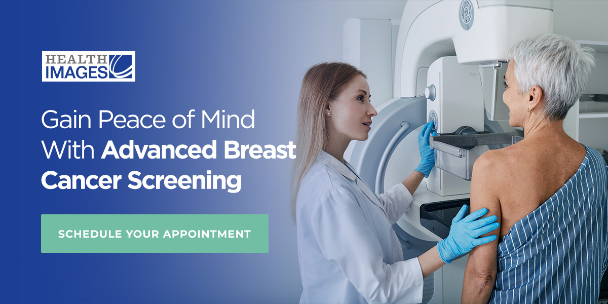
Gain Peace of Mind With Advanced Breast Cancer Screening
An ABUS exam enhances breast cancer screening for early detection and greater peace of mind. It’s an effective supplement to mammograms if you have dense breasts because it can reveal findings that may not appear on standard mammogram images.
We offer ABUS screenings to help you take a proactive approach to your health. Our compassionate team is dedicated to providing a warm, comfortable experience with accurate results. Learn more about our services or request an ABUS screening today.
 If you have dense breasts, supplemental breast cancer screening is an excellent way to gain peace of mind and be proactive about your health. Supplemental screening methods, such as an automated breast ultrasound (ABUS), can reveal abnormalities that dense tissue may hide on a standard mammogram. While annual mammograms remain the most important breast cancer screening tool, supplemental screening can increase your chances of early detection.
 Breast density refers to the composition of the breast, or how much fatty tissue and connective tissue are present. Dense breasts have more glandular and fibrous tissue, and non-dense breasts are made up of more fatty tissue. Fibrous tissue provides structural support, and glandular tissue consists of milk glands and the ducts that transport milk following a pregnancy. These tissues are what give breasts their particular shape and firmness, and their relative amount varies by person.
Dense and non-dense breasts are completely normal, but they can impact how tissue and cancer cells appear on screens.
Breast density refers to the composition of the breast, or how much fatty tissue and connective tissue are present. Dense breasts have more glandular and fibrous tissue, and non-dense breasts are made up of more fatty tissue. Fibrous tissue provides structural support, and glandular tissue consists of milk glands and the ducts that transport milk following a pregnancy. These tissues are what give breasts their particular shape and firmness, and their relative amount varies by person.
Dense and non-dense breasts are completely normal, but they can impact how tissue and cancer cells appear on screens.
 The Food and Drug Administration (FDA) requires mammography providers to inform each patient whether their breasts are “dense” or “not dense” in their mammogram reports. For a more detailed explanation of your breast density, you can ask your provider for your full mammography report. While simplified reports categorize density as dense or not dense, a detailed report reveals which of the following breast imaging reporting and data system (BI-RADS) categories your breasts are part of:
 The following factors can impact breast density:
 Supplemental screening options are available for women with dense breasts, such as an MRI, handheld ultrasound or an automated breast ultrasound. An ABUS ultrasound is a supplemental breast cancer screening test for women with dense breasts. It creates a 3D image of the breast with ultrasound technology, catching abnormalities that may not appear on a mammogram. This technology uses an automated scanner and sound waves that bounce off different tissues. The scanner converts the echoes into electronic signals, and a computer processes these signals into detailed 3D images.
 Use the following tips to prepare for an ABUS exam:
 An ABUS screening is noninvasive, taking about 15-20 minutes and involving the following steps:
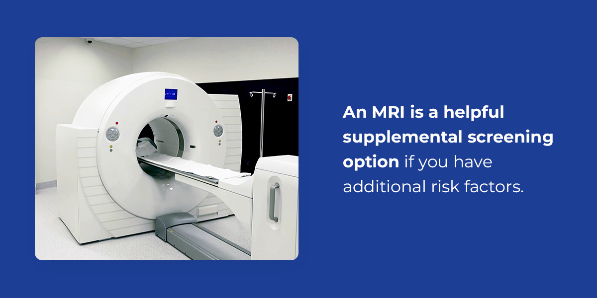 In some cases, your provider may recommend an MRI instead of an ABUS. An MRI is a helpful supplemental screening option if you have additional risk factors, such as:
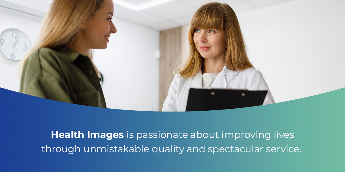 Health Images is passionate about improving lives through unmistakable quality and spectacular service. Trusting us with your ABUS screening needs offers the following advantages: