- Screening mammograms: These are a part of routine medical care. The American Cancer Society recommends annual screening mammograms for women 45-54 years old and advises that women should have the option to begin screening as early as 40.
- Diagnostic mammograms: This type of mammogram occurs more variably. Your doctor may recommend a diagnostic mammogram if a manual exam or screening has revealed any changes or potential indicators of breast cancer. Because diagnostic mammograms also provide more in-depth imaging, your doctor may recommend one if you have dense breast tissue.
What are mammograms?
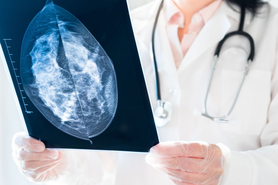
A mammogram is a type of low-dose x-ray that examines breast tissue for potential signs of cancer. Low-density tissue like fat appears translucent in the x-ray images, while higher-density tissues like glands or tumors appear white. Doctors can examine changes in the tissue or lumps too small to detect during a manual examination by studying these images.Digital mammograms vs. regular mammograms
Another way to categorize mammograms is by how they record their data. Traditional mammograms produce the type of x-rays most people are familiar with, where doctors read and store the images on film. Regular mammograms are subject to potential flaws in the film and the limited perspective of each image, which can sometimes lead to inaccurate diagnoses and false positives. Digital mammograms record their images on a computer rather than on film, allowing your doctor to easily share them for consultations. Digital x-rays also produce better picture quality with higher levels of detail. The images can be enhanced, manipulated or magnified, making evaluations easier and minimizing the chances of a false-positive diagnosis. While imaging facilities require FDA approval to offer digital mammograms, the process for both methods is identical for patients. Health Images exclusively uses digital mammography, giving you the clearest imagery possible.
What are the benefits of mammograms?
In addition to diagnostic screenings’ obvious utility in identifying possible signs of breast cancer, screening mammograms offer several benefits as well:- Early detection: Screening mammograms can detect signs of breast cancer before the breast tissue begins producing noticeable manifestations or symptoms. This early detection allows more immediate treatment, which may catch cancer before it can spread.
- Improved survival rates: Regular breast cancer screenings can reduce breast cancer’s mortality risk by up to 49%.
- Lower mastectomy rates: Catching breast cancer in its early stages can improve your chances of successful treatment without requiring a mastectomy.
Bilateral mammograms vs. 3D mammograms
When you’re deciding how you want the procedure performed, you have two types of mammograms to choose from. The bilateral and 3D mammogram processes are nearly identical from the patient perspective. Both methods function on one breast at a time, compressing the tissue with a paddle to maximize the x-ray image quality with minimal radiation. While a 3D mammogram may take slightly longer, both methods typically take about 10 minutes. While bilateral and 3D mammograms are functionally the same for the patient, there are significant differences for the radiologists and doctors who interpret the information.Bilateral mammography
Bilateral mammograms represent the standard or traditional type of mammography. In these, the mammogram machine x-rays the breast tissue from a top and side view. Since a bilateral screening mammogram only shows the breast tissue from two angles, there’s little compensation for overlap. In addition to fat, breasts contain supportive tissue, blood vessels, ligaments, milk-processing glands and ducts. When these overlap on an x-ray, they can obscure tumors and other signs of breast cancer, leading to false negatives. The overlap can also appear to be a tumor itself, leading to a false positive.3D mammography
While bilateral mammograms take one image from the top and one image from the side, 3D mammography takes many.
A 3D mammogram machine moves in an arc to take multiple low-dose x-rays of the breast from different angles. Those images combine into a picture composed of different views, producing a three-dimensional model for doctors to review. Rather than trying to interpret overlapping tissues from a single perspective, they can shift to a different view to identify the parts of what they see.
This more detailed image makes it easier to find breast abnormalities even within dense surrounding tissue. It also allows radiologists to understand the location, shape and size of those abnormalities, helping them identify which are benign and which are potential tumors.
3D mammography’s high-quality images and comprehensive modeling offer several benefits:
- Improved breast cancer detection: 3D mammography lets doctors and radiologists view the breast from multiple angles. In doing so, they can spot cancers that dense breast tissue might otherwise obscure. This method also helps them find small cancers earlier. Earlier detection can lead to quicker treatment and smaller removal sites, with lower mastectomy risk.
- Fewer false positives: The mammogram’s three-dimensional modeling allows doctors to view the breast at different angles, helping them rule out areas of overlap.
- Reduced callbacks and follow-up imaging: A more comprehensive array of images offers a greater opportunity for doctors to interpret the x-rays correctly. With sufficient data from the initial mammogram, they can draw their conclusions more confidently.
- Reduced patient anxiety: Greater confidence from the doctor translates to greater patient confidence. As the odds of callbacks and false positives go down, anxiety decreases.
Frequently asked questions
For more information on the mammography process, here are a few frequently asked questions.Is tomosynthesis the same as 3D mammography?
Yes, it is. 3D mammography is also known as digital breast tomosynthesis (DBT). While DBT produces its images in a way similar to computed tomography (CT) scanners, DBT uses far fewer x-ray beams than CT scanners do.Are 3D mammograms better for dense breasts?
Dense breast tissue — the breasts’ milk glands, supportive tissue and milk ducts — can have the same obscuring effect as overlap in bilateral mammograms. 3D mammography’s modeling lets doctors spot abnormalities even in dense tissue more accurately.Is the radiation from mammogram machines dangerous?
With lower levels than either CT scans or regular chest x-rays, radiation from mammograms is within medical guidelines and completely safe.Request a screening mammogram with Health Images
At Health Images, we believe in doing the right thing in the right way for the right reasons. We’re committed to ensuring your comfort while providing top-quality care, and our all-digital mammography and electronic medical record delivery mean your results reach your referring physician in hours. You can get in touch with us at a location near you to request your screening mammogram today.Call To Schedule Your Appointment
Sources (cited):- https://www.fda.gov/radiation-emitting-products/mqsa-insights/dbt-accreditation-its-here
- https://www.cancer.org/healthy/find-cancer-early/american-cancer-society-guidelines-for-the-early-detection-of-cancer.html
- https://www.healthimages.com/diagnostic-mammogram-vs-screening-mammogram/
- https://www.cancer.gov/types/breast/mammograms-fact-sheet
- https://pubs.rsna.org/doi/10.1148/radiol.2021203935?_ga=2.200402414.1709253957.1618236126-143523632.1570560266
- https://bmccancer.biomedcentral.com/articles/10.1186/s12885-021-07917-2
- https://www.healthline.com/health/how-long-does-a-mammogram-take#how-long-does-it-take
- https://www.healthimages.com/services/mammography/
- https://www.healthimages.com/breast-density-on-mammograms/
- https://www.healthimages.com/6-myths-about-mammograms-busted/
- https://www.healthimages.com/locations/?service=mammography