Imaging tests help diagnose medical conditions. Whether an x-ray to assess bone fractures or a CT scan to visualize internal organs, these tests provide valuable information that helps healthcare providers make informed decisions about patient care.
You may have heard about the association between medical imaging tests and radiation, and it’s natural to have questions regarding how much radiation is in a CT scan or x-ray. We’ll cover the types of radiation and imaging tests, as well as the amount of radiation you receive from imaging tests.
Types of radiation
Radiation is energy that travels in waves or particles and can come from various sources. The two main types of radiation related to medical imaging tests are ionizing and non-ionizing radiation.
Ionizing radiation
Ionizing radiation uses high-energy waves to remove tightly bound electrons from atoms. When electrons are lost, the atom or molecule becomes electrically charged, called an ion. This process can potentially cause damage to living tissues.
Ionizing radiation is commonly used in imaging tests such as x-rays, CT scans and nuclear medicine scans. The risks associated with ionizing radiation are typically low and carefully regulated in medical imaging tests to ensure patient safety.
Non-ionizing radiation
Non-ionizing radiation uses lower-energy waves and lacks the ability to remove tightly bound electrons from atoms. This type of radiation is used in imaging tests such as MRI scans and ultrasounds. Non-ionizing radiation is generally considered safe, as it does not carry the same risk of cellular damage associated with ionizing radiation.
Background radiation
In addition to being used in medical imaging tests, radiation exists naturally in our environment. The levels of natural radiation exposure vary depending on geographical location, altitude and lifestyle choices.
The majority of background radiation occurs from minerals in the ground, soil and water. Cosmic radiation also contributes to background radiation.
Radiation in daily life
Radiation is a natural part of our environment, and we are exposed to it daily. Here are some common examples of background radiation:
Cosmic radiation: Cosmic radiation originates from outer space and includes high-energy particles from the sun and stars. It is constantly bombarding the Earth’s atmosphere.
Radon gas: Radon is a naturally occurring radioactive gas produced by the decay of uranium and radium in soil and rocks. It can seep into buildings through cracks in the foundation and accumulate indoors, especially in poorly ventilated areas.
Terrestrial radiation: Terrestrial radiation comes from naturally occurring radioactive materials in the Earth’s crust, such as uranium, thorium and potassium.
Consumer products: Certain products, such as smoke detectors, ceramic and granite tiles, and some electronic devices, contain small amounts of radioactive materials and can contribute to background radiation. However, the radiation levels from these products are generally very low and not considered a significant health risk.
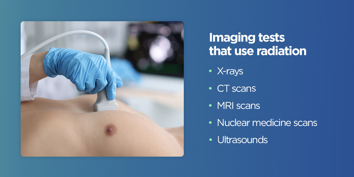
Imaging tests that use radiation
Each imaging test has strengths, limitations and appropriate uses. Healthcare providers carefully determine the most appropriate test for each individual. The following imaging tests use radiation:
X-rays: X-rays use a small amount of ionizing radiation to create images of bones, teeth and specific organs. They are used to diagnose conditions such as fractures, infections and lung diseases. Mammography is an example of an x-ray procedure. Mammography uses an x-ray system to see inside the breasts.
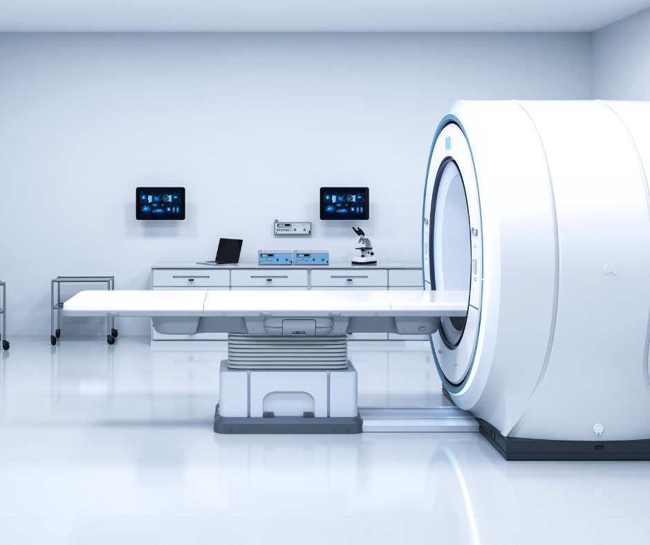
Computed tomography (CT) scans: Also known as CAT scans, CT scans provide detailed cross-sectional images of the body. They use x-rays and advanced computer technology to create clear images of internal structures. CT scans are often used to diagnose conditions such as cancer, infections and trauma-related injuries.
Magnetic resonance imaging (MRI): MRI scans use powerful magnets and radio waves to generate detailed images of the body. They help visualize soft tissues like the brain and muscles. MRI scans do not involve ionizing radiation, making them a safer option for populations such as pregnant women and children.
Nuclear medicine scans: Nuclear scans involve using small amounts of radioactive substances, known as radiopharmaceuticals, to visualize functional changes in the body. Nuclear medicine scans can help diagnose conditions such as cancer, heart disease and bone disorders.
Ultrasounds: Ultrasounds are noninvasive procedures that use sound waves to create images of the body’s internal structures.
Radiation in imaging tests
The amount of radiation you can receive from an imaging test is carefully regulated. Radiology departments follow strict protocols to ensure patient safety. These protocols include using the lowest possible radiation dose that still provides adequate image quality, using shielding devices to protect sensitive areas of the body and following established guidelines for radiation safety.
Explore potential radiation exposure from the following imaging tests:
Do CT scans use radiation? CT scans generally involve higher radiation doses than other imaging tests. The radiation exposure can range from 2 to 10 millisieverts (mSv) per dose, depending on the type of CT scan and the body part being imaged. Some complex CT scans or repeated scans may result in higher radiation exposure.
Do x-rays use radiation? The radiation dose from an x-ray varies based on the body part. A hand or foot x-ray exposes you to less than 0.001 mSv per dose, while receiving an x-ray of your large intestine exposes you to 6 mSv per scan.
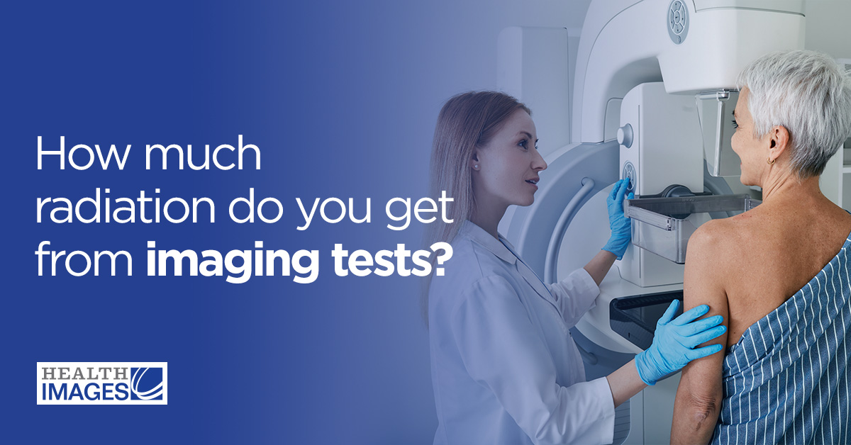
Do mammograms use radiation? The radiation dose from a mammogram is generally low, averaging around 0.4 mSv for a standard mammogram with two views of each breast. However, the radiation dose can vary depending on the type of mammogram (digital or analog), the breast tissue density and the imaging technique used.
To put these radiation doses into perspective, the average annual background radiation dose from natural sources in the United States is estimated to be around 3 mSv per year, although this can vary depending on geographic location.
Is there a cancer risk from imaging tests?
The risk of developing cancer from imaging tests is generally considered low. However, patients who receive frequent or repetitive imaging tests should consider cumulative radiation exposure over time. For example, if you receive multiple imaging tests with ionizing radiation over your lifetime, you may be at a slightly increased risk of developing cancer compared to the general population.
Your healthcare provider can provide personalized information and guidance based on your situation. You’ll want to keep track of your imaging history and inform your healthcare provider of any past imaging tests you have had so that they can take your cumulative radiation exposure into account when determining the need for future imaging tests.
Benefits and risks of imaging tests
Like any medical procedure, imaging tests have benefits and risks. Imaging tests provide valuable information that helps healthcare providers make accurate diagnoses, guide treatment plans and monitor the effectiveness of interventions. They can also reduce the need for more invasive procedures by providing detailed images of the body’s internal structures.
One of the main risks associated with imaging tests is the use of ionizing radiation. However, the risks are generally considered low, especially considering modern imaging techniques that use lower doses of radiation. Other risks may include allergic reactions to contrast agents used in certain tests, discomfort or claustrophobia during some procedures, and the possibility of false positive or false negative results.
Safeguard your health during imaging tests with Health Images
If you need an imaging test, work with qualified healthcare professionals who use modern and well-maintained imaging equipment. Providers like Health Images prioritize patient safety and follow established protocols to ensure that radiation doses are kept as low as reasonably achievable while obtaining accurate diagnostic images.
When you work with experienced technologists, you can have peace of mind knowing that your imaging tests are conducted with safety in mind. With proper medical guidance and appropriate utilization of imaging tests, you can make informed decisions to safeguard your health.
 Imaging tests help diagnose medical conditions. Whether an x-ray to assess bone fractures or a CT scan to visualize internal organs, these tests provide valuable information that helps healthcare providers make informed decisions about patient care.
You may have heard about the association between medical imaging tests and radiation, and it’s natural to have questions regarding how much radiation is in a CT scan or x-ray. We’ll cover the types of radiation and imaging tests, as well as the amount of radiation you receive from imaging tests.
Imaging tests help diagnose medical conditions. Whether an x-ray to assess bone fractures or a CT scan to visualize internal organs, these tests provide valuable information that helps healthcare providers make informed decisions about patient care.
You may have heard about the association between medical imaging tests and radiation, and it’s natural to have questions regarding how much radiation is in a CT scan or x-ray. We’ll cover the types of radiation and imaging tests, as well as the amount of radiation you receive from imaging tests.
 Each imaging test has strengths, limitations and appropriate uses. Healthcare providers carefully determine the most appropriate test for each individual. The following imaging tests use radiation:
 If you need an imaging test, work with qualified healthcare professionals who use modern and well-maintained imaging equipment. Providers like Health Images prioritize patient safety and follow established protocols to ensure that radiation doses are kept as low as reasonably achievable while obtaining accurate diagnostic images.
When you work with experienced technologists, you can have peace of mind knowing that your imaging tests are conducted with safety in mind. With proper medical guidance and appropriate utilization of imaging tests, you can make informed decisions to safeguard your health.
If you need an imaging test, work with qualified healthcare professionals who use modern and well-maintained imaging equipment. Providers like Health Images prioritize patient safety and follow established protocols to ensure that radiation doses are kept as low as reasonably achievable while obtaining accurate diagnostic images.
When you work with experienced technologists, you can have peace of mind knowing that your imaging tests are conducted with safety in mind. With proper medical guidance and appropriate utilization of imaging tests, you can make informed decisions to safeguard your health.