If you’re experiencing frequent pain and inflammation in your bones, joints or muscles, you might be dealing with a musculoskeletal disorder. You’re not alone — these disorders can be quite common, especially as you get older or perform repetitive movements.
Like many medical conditions, treating a musculoskeletal disorder requires an accurate diagnosis. Luckily, there are many imaging tests available to help your medical provider identify the issue. From there, they can help you determine the right treatment path to alleviate pain and improve strength and movement.
If you’re seeking relief from a bone- or muscle-related condition, continue reading for a list of musculoskeletal disorders, causes, symptoms, imaging techniques and treatment methods.
What is a musculoskeletal disorder?
A musculoskeletal disorder (MSD) is a broad term describing an injury or impairment in one of these areas:
Muscles
Tendons
Nerves
Joints
Cartilage
Spinal discs
Some common types of MSDs include:
Bone fractures: Overuse, disease and trauma can weaken the bones, sometimes to the point of a partial or full break.
Carpal tunnel syndrome: Carpal tunnel syndrome is a pinched nerve in the wrist causing tingling or numbness in the hand. It typically stems from excessive or repetitive hand and wrist use.
Osteoarthritis: One of the most common types of arthritis, osteoarthritis occurs when bone cartilage gradually wears down. This can cause pain and inflammation in the lower back, neck, hands, hips or knees.
Rheumatoid arthritis: Rheumatoid arthritis is another common type of arthritis where the immune system attacks its own cells. It usually inflames the joint cartilage but can spread to other organs as well.
Tendinitis: Tendinitis is the inflammation of a tendon. It often affects the wrists, elbows, shoulders and ankles.
Symptoms of MSDs
Because MSDs encompass a wide range of conditions, people can experience a variety of symptoms. Pain is one of the earliest indicators, but it varies by person and condition. Someone might experience pain in a particular region, while another might have pain throughout the body. Some common MSD symptoms include:
Fatigue
Recurring aches, pain and discomfort with varying severity
Stiff or weak muscles
Tingling and numbness
Burning sensation
Reduced strength and movement
Swelling and inflammation
Muscle spasms
Discoloration or bruising
What causes MSDs?
MSDs often stem from repetitive movement or workplace injuries. There may be a genetic factor involved in conditions like arthritis. However, MSDs are typically related to the following factors:
Aging
Poor posture when standing, walking, sitting or lifting
Routinely bending, twisting or lifting heavy objects
When an MSD develops, factors like poor dietary habits, obesity and a sedentary lifestyle can aggravate the condition.
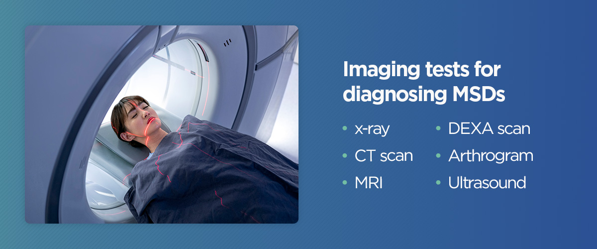
Imaging tests for diagnosing MSDs
Your healthcare provider may order one or more imaging tests to examine your bones, joints and muscles more closely. The type of imaging test suggested will primarily depend on what they believe the condition is. Below are some imaging techniques for common musculoskeletal disorders.
1. X-ray
An x-ray is usually the first type of imaging a medical provider will order when they believe a patient has an MSD. X-rays use electromagnetic waves to produce images.
The picture’s clarity depends on how well the waves pass through the bone or organ. Bones tend to absorb these waves more effectively than soft tissues and muscles. Your healthcare provider might order a stress x-ray to determine the amount of damage to a joint, which involves moving or positioning the joint at multiple angles.
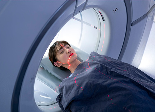
2. CT scan
A computerized tomography (CT) scan uses multiple x-ray views captured from different angles. Modern scanners can provide 3D images of structures.
During a CT scan, you’ll lie flat on a table that moves through a donut-shaped tube. The tube will move around your body, collecting images from various angles. In some cases, contrast dye is used to make irregularities more visible.
3. MRI
Magnetic resonance imaging (MRI) involves magnetic fields and radio waves to collect images within the body. You’ll lie still while moving through a long, tube-shaped machine. Some patients find enclosed MRI tubes anxiety-inducing and opt for open MRI scanners instead.
An MRI generates 3D images resembling cross-sections inside of the body. Unlike an x-ray, it can capture muscle and soft tissue images clearly. However, this technique isn’t suitable for people with implants, pacemakers or metal plates since it uses magnets.
4. DEXA scan
A dual-energy x-ray absorptiometry (DEXA) scan measures bone density and mass with x-ray imaging. It’s often used to diagnose conditions causing bone loss, like osteoporosis. DEXA scans can also assess fat and muscle mass.
A DEXA scan normally takes up to 30 minutes. You lie flat on a table while a scanner passes over you from above. It uses two types of x-ray beams to capture images.
5. Arthrogram
An MR/CT arthrogram takes images of the inside of a joint. A technologist injects contrast dye into the joint, then moves the joint in different positions while taking pictures. An arthrogram allows them to identify joint-related issues that standard imaging might have missed. Doctors often order arthrograms for patients with unexplained joint problems, like loss of motion or painful movement.
A possible concern with arthrograms is that some patients might be allergic to the dye. If you know you’re allergic to any common ingredients in the dye or you’ve previously had a reaction to contrast dye, let your medical provider know. They may perform a different type of imaging test instead.
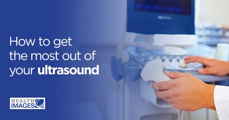
6. Ultrasound
Ultrasounds and sonograms use sound waves to take images of the inside of the body. They’re great at capturing soft tissues, muscles and ligaments. An ultrasound is especially helpful for pinpointing signs of inflammation and swelling. Many people associate ultrasounds and sonograms with pregnancy, but they can also diagnose MSDs.
Musculoskeletal disorder treatment options
Your doctor will recommend a treatment plan upon diagnosing an MSD. Depending on the severity, this can include:
Over-the-counter medications
Moderate exercise
Physical therapy or occupational therapy
Splints or wrist supports
You can also help prevent MSDs by developing the following habits:
Stretching and exercising regularly for strong bones, muscles and joints
Stopping or changing positions when an activity causes pain
Request an imaging test at Health Images
If you think you might have a musculoskeletal disorder, our team at Health Images in Colorado can assist you. With our state-of-the-art equipment, personalized service, knowledgeable staff and world-class diagnostic imaging, we can provide the customized care and accurate results needed to address the problem.
Whatever type of imaging technique you require, we’ll guide you through the process to ensure you feel comfortable and relaxed. Find your nearest Health Images location to request an x-ray, MRI, CT scan or other diagnostic imaging today.
 If you’re experiencing frequent pain and inflammation in your bones, joints or muscles, you might be dealing with a musculoskeletal disorder. You’re not alone — these disorders can be quite common, especially as you get older or perform repetitive movements.
Like many medical conditions, treating a musculoskeletal disorder requires an accurate diagnosis. Luckily, there are many imaging tests available to help your medical provider identify the issue. From there, they can help you determine the right treatment path to alleviate pain and improve strength and movement.
If you’re seeking relief from a bone- or muscle-related condition, continue reading for a list of musculoskeletal disorders, causes, symptoms, imaging techniques and treatment methods.
If you’re experiencing frequent pain and inflammation in your bones, joints or muscles, you might be dealing with a musculoskeletal disorder. You’re not alone — these disorders can be quite common, especially as you get older or perform repetitive movements.
Like many medical conditions, treating a musculoskeletal disorder requires an accurate diagnosis. Luckily, there are many imaging tests available to help your medical provider identify the issue. From there, they can help you determine the right treatment path to alleviate pain and improve strength and movement.
If you’re seeking relief from a bone- or muscle-related condition, continue reading for a list of musculoskeletal disorders, causes, symptoms, imaging techniques and treatment methods.
 Your healthcare provider may order one or more imaging tests to examine your bones, joints and muscles more closely. The type of imaging test suggested will primarily depend on what they believe the condition is. Below are some imaging techniques for common musculoskeletal disorders.