Women’s imaging involves screening and diagnostic testing to improve women’s health and reduce the risk of disease development. Since women can experience unique medical conditions — such as menopause, pregnancy and reproductive issues — they need specialized treatment and testing throughout each stage of life. These tests provide immediate, accurate results for medical professionals to treat their patients accordingly. Explore the various women’s health care imaging testing available today.
Women’s Health Imaging Benefits
Imaging for women provides the following advantages for patients and medical professionals.
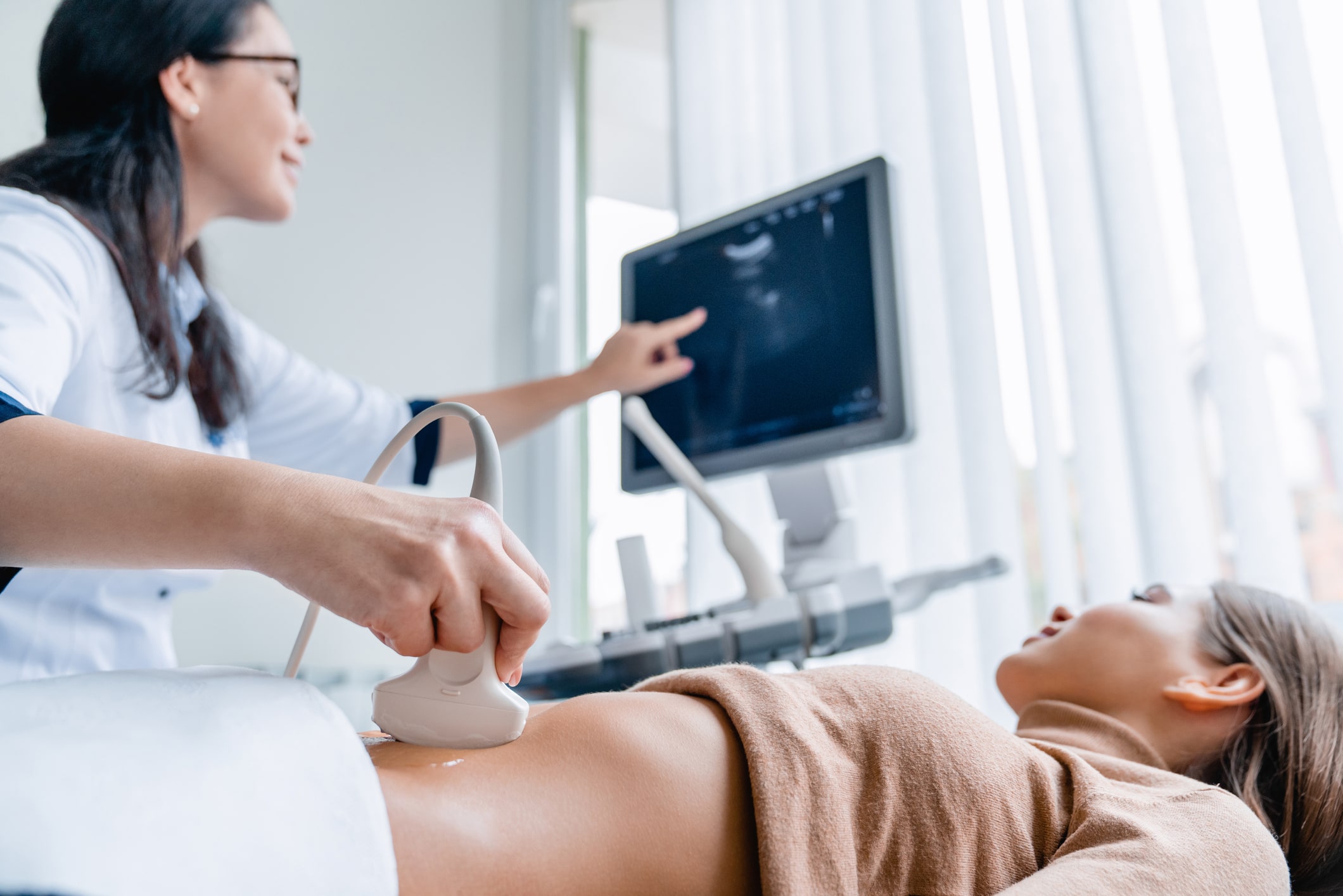
Track progress during pregnancy: Technologists perform ultrasounds on pregnant women to discover how their babies are developing. After reviewing the sonogram, the doctor can also diagnose any issues that arise so the baby can continue to grow healthy and strong in the womb.
Obtain immediate results: Medical professionals can see the results of these diagnostic tests as soon as possible to diagnose their patients’ symptoms accurately and create the appropriate treatment plan.
Get a clear picture: A women’s imaging test provides a conclusive view of a specific area of the body. Doctors can use these tests to assess a patient’s exact symptoms and decide on the best treatment options.
Improve patients’ well-being: Diagnostic imaging for women provides early detection of cancer and other diseases, so doctors can catch them as soon as possible. Treatment is most effective during the earliest stages of the disease’s development. Mammograms also reduce the need for a mastectomy, or full breast removal, during cancer recovery.
Customize screening: Women’s imaging testing allows doctors to run specific tests to diagnose common issues in women of all ages.
Track current disease treatment: Medical imaging for women can inform doctors of their patients’ treatment progress to analyze effectiveness.
Limit future health issues: Screening and diagnostic tests for women advise medical professionals of underlying health issues they’ll need to address in their patients to prevent the development of diseases and other medical conditions.
Types of Diagnostic Imaging for Women
A women’s imaging center offers the following screening and diagnostic tests.
1. Mammogram
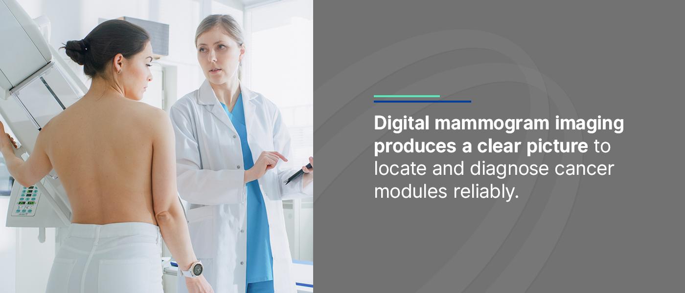
A mammogram involves taking x-ray pictures of a patient’s breasts. These images help doctors catch early signs of breast cancer, such as abnormal breast tissue changes and lumps that are too small for the patient to feel. When they detect these symptoms, a doctor can begin treatment as soon as possible before they develop further. Medical professionals recommend scheduling an annual women’s mammogram starting at age 40.
Digital mammogram imaging produces a clear picture to locate and diagnose cancer modules reliably. Medical professionals typically use two primary types of digital mammograms.
Screening mammography: Technologists perform annual screening mammograms to look for unsuspected breast cancer, even if the woman doesn’t display any signs or symptoms. This imaging testing helps prevent breast cancer development and gives the patient peace of mind.
Diagnostic mammography: A doctor may recommend a patient for a diagnostic mammogram if a screening exam indicates any signs or symptoms of breast cancer. The patient may also get this diagnostic testing if abnormal symptoms, such as nipple discharge, a palpable lump or skin dimpling, are present.
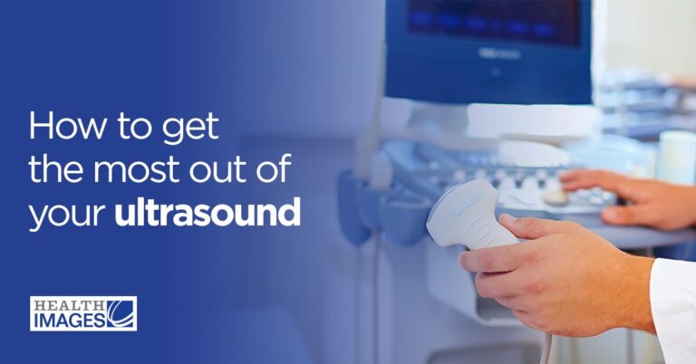
An ultrasound features high-frequency sound waves that provide real-time images inside a patient’s body, including the structure of internal organs and blood flow. This imaging test produces an accurate picture because the sound waves can travel through fluid and soft tissue, but bounce back when they hit denser surfaces. It’s also a safe testing method for pregnant people because it doesn’t use radiation.
When do medical professionals perform ultrasounds?
Regular pregnancy appointments: An ultrasound can help technologists identify the fetus’ position in the womb, monitor growth and determine the baby’s sex.
Gynecological exams: Doctors can also use ultrasounds to detect ovarian and cervical cancer.
Mammogram follow-ups: If a woman’s mammogram doesn’t provide enough information, the doctor may recommend an ultrasound to assess breast tissue abnormalities and other health conditions.
A doctor can use an ultrasound to determine whether a lump in the breast, ovary or cervix is a benign cyst or the potential development of cancer. It can also help medical professionals target the right place to perform a biopsy.
3. Breast MRI Scans
A breast MRI scan uses radiation to provide a detailed image of the breast by looking at it from various angles, allowing doctors to examine tissue type and density. Medical professionals will recommend a breast MRI if an ultrasound or mammogram test brings up insufficient details to provide a conclusive diagnosis. They may also perform fetal MRI scans for pregnant people if they need more precise information after the ultrasound.
Like a mammogram, patients who are at a higher risk of breast cancer may get a breast MRI scan as a screening tool. Instead of replacing a mammogram or ultrasound, this diagnostic testing serves as an additional tool to provide accurate diagnosis and treatment for patients.
4. Bone Density Scan
Medical professionals use a bone density scan, or bone mineral density testing, to measure the amount of calcium per square centimeter within a patient’s bones. As a result, doctors can determine their patients’ chances of fracture and treat women with a lower bone density and lack of minerals. Medical professionals can also use bone density scans to track a medication’s effectiveness for treating existing bone conditions in their patients.
Since women typically have thinner bones than men, they’re usually more prone to osteoporosis and osteopenia. When their estrogen declines as they go through menopause, they’ll need to have their bone density checked. This imaging test determines a patient’s bone fragility and what precautions to take to prevent fracture. Doctors usually suggest that women get bone density scans at age 65 or older, or 50 years old if they’ve previously broken a bone.
5. DEXA
A doctor will usually recommend a full BodyLogic™ DEXA Body Composition scan to measure fat mass, lean mass and bone density for a detailed image of the patient’s body. Athletes typically use this body composition scan to help them decide on the best training program based on their specific muscles and bones. This information helps medical professionals accurately assess their patient’s health to provide the best treatment plan. A DEXA scan provides immediate results so the doctor can quickly analyze and report the patient’s health.
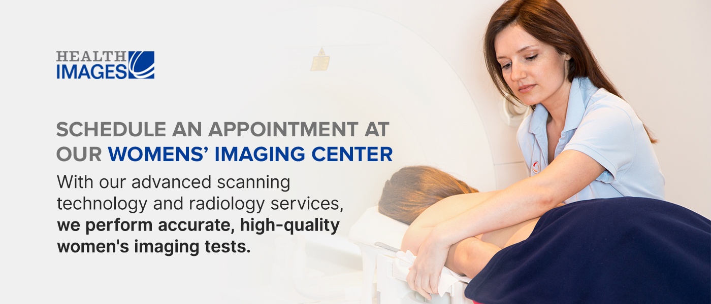
Schedule an Appointment at Our Women’s Imaging Centers
Health Images offers various services for our female patients in the Denver area. With our advanced scanning technology and radiology services, we perform accurate, high-quality women’s imaging tests. Our professional technologists combine a pleasant imaging experience with passionate care to enhance patient health. Book an appointment with us online or over the phone today to get screening or diagnostic testing.
Sources:
 Women’s imaging involves screening and diagnostic testing to improve women’s health and reduce the risk of disease development. Since women can experience unique medical conditions — such as menopause, pregnancy and reproductive issues — they need specialized treatment and testing throughout each stage of life. These tests provide immediate, accurate results for medical professionals to treat their patients accordingly. Explore the various women’s health care imaging testing available today.
 Digital mammogram imaging produces a clear picture to locate and diagnose cancer modules reliably. Medical professionals typically use two primary types of digital mammograms.
 Sources: