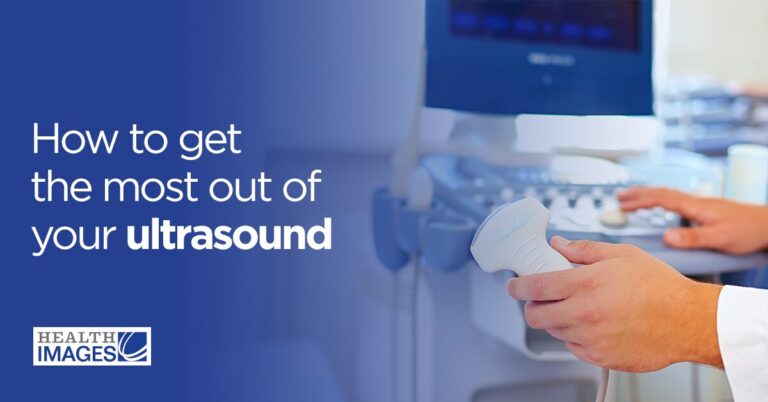
 You may have heard the words “sonogram” and “ultrasound” used interchangeably, and it’s easy to get the two confused. What do these terms mean exactly, and how are they different? In this post, we’ll clarify the meaning of these easy-to-confuse words and provide some information about both. If you want to learn more about imaging procedures, we’ll be happy to assist you at Health Images.
Jump to Sections:
You may have heard the words “sonogram” and “ultrasound” used interchangeably, and it’s easy to get the two confused. What do these terms mean exactly, and how are they different? In this post, we’ll clarify the meaning of these easy-to-confuse words and provide some information about both. If you want to learn more about imaging procedures, we’ll be happy to assist you at Health Images.
Jump to Sections:
- Are Ultrasound and Sonogram the Same Thing?
- What Is a Sonogram?
- How Does a Sonogram Work?
- What Is a Sonogram Used For?
- What Is an Ultrasound?
- How Does an Ultrasound Work?
- What Is an Ultrasound Used For?
- What’s the Difference Between a Sonogram and an Ultrasound?
- How Are Sonograms and Ultrasounds Similar?
Are Ultrasound and Sonogram the Same Thing?
Although “ultrasound” and “sonogram” are both used to refer to the same exam, they have different meanings. We’ll explore the differences and similarities between these terms below and explain how they work.What Is a Sonogram?
A sonogram is an image produced by an ultrasound. In other words, it’s not the procedure itself but the product. For example, many parents can recall the thrilling moment they saw their baby on the ultrasound monitor and were then given the printed sonogram to take home. In this instance, the sonogram is the black, white and gray image that shows the fetus in the uterus. You may also hear the word “sonography” used in reference to the ultrasound exam. Sonography refers to the practice of using an ultrasound machine to create a sonogram, and the sonographer is the skilled technologist who operates the equipment.How Does a Sonogram Work?
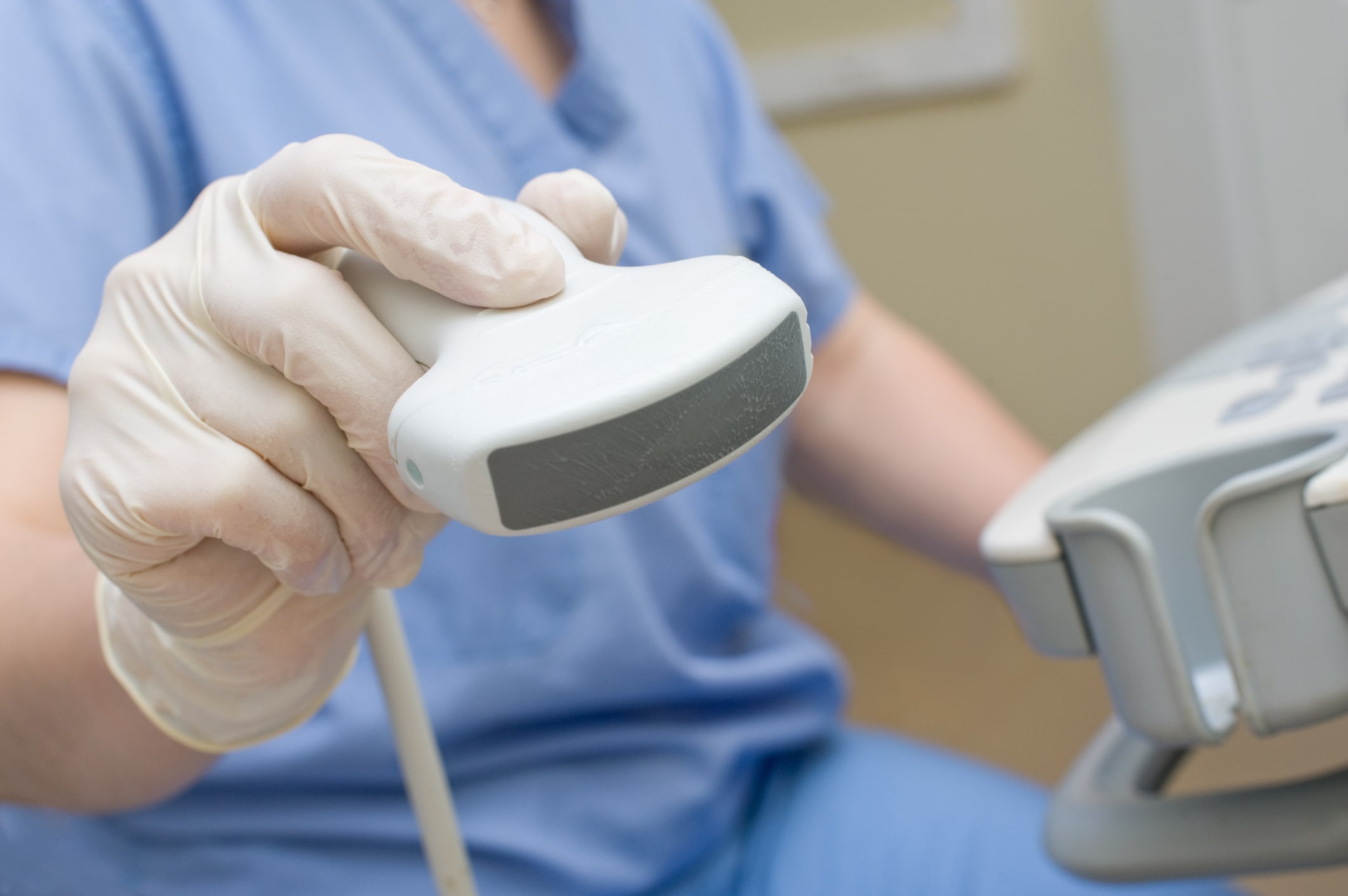
A sonogram is produced by sound waves. During an ultrasound, the technologist will send sound waves to the part of the body under examination using a wand-shaped device called a transducer. The sound waves will bounce back once they contact the tissues being studied, travel through the transducer and enter the ultrasound computer. The computer will process the sound waves and turn them into an image, which the patient and technologist can view on the monitor. Sound waves will create images only when they can reflect from a surface. For example, sound waves travel easily through fluids and will not bounce off of water, urine or other liquids. They will, therefore, appear black on a sonogram. When sound waves hit a tissue, they reflect back into the ultrasound machine and transform into a white or gray image, depending on the intensity of the sound wave. Extremely dense tissues such as bones and kidney stones produce echoes easily and appear bright white on a sonogram.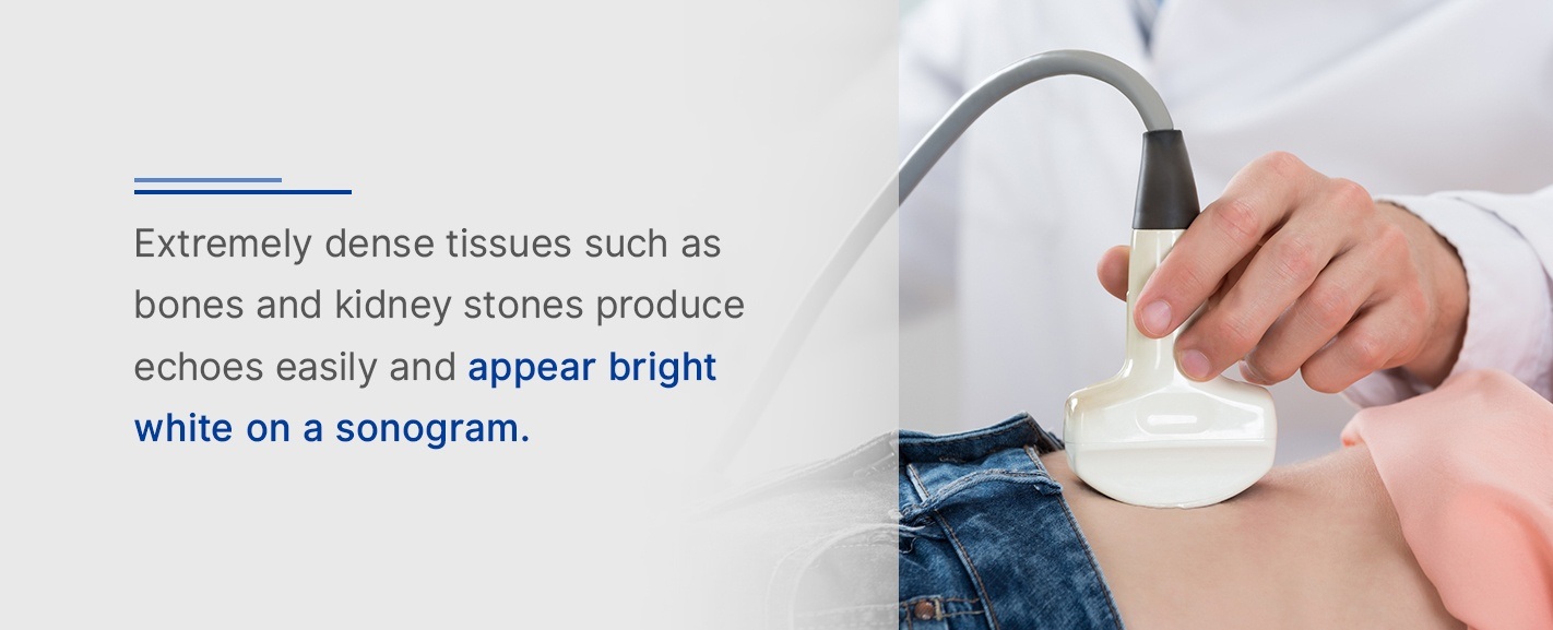
What Is a Sonogram Used For?
As we’ll discuss more below, sonograms help doctors evaluate organs for infections, damage or disease. Pregnant women may get ultrasounds to generate sonograms of the fetus. This process allows a doctor to check a baby’s development and health.What Is an Ultrasound?
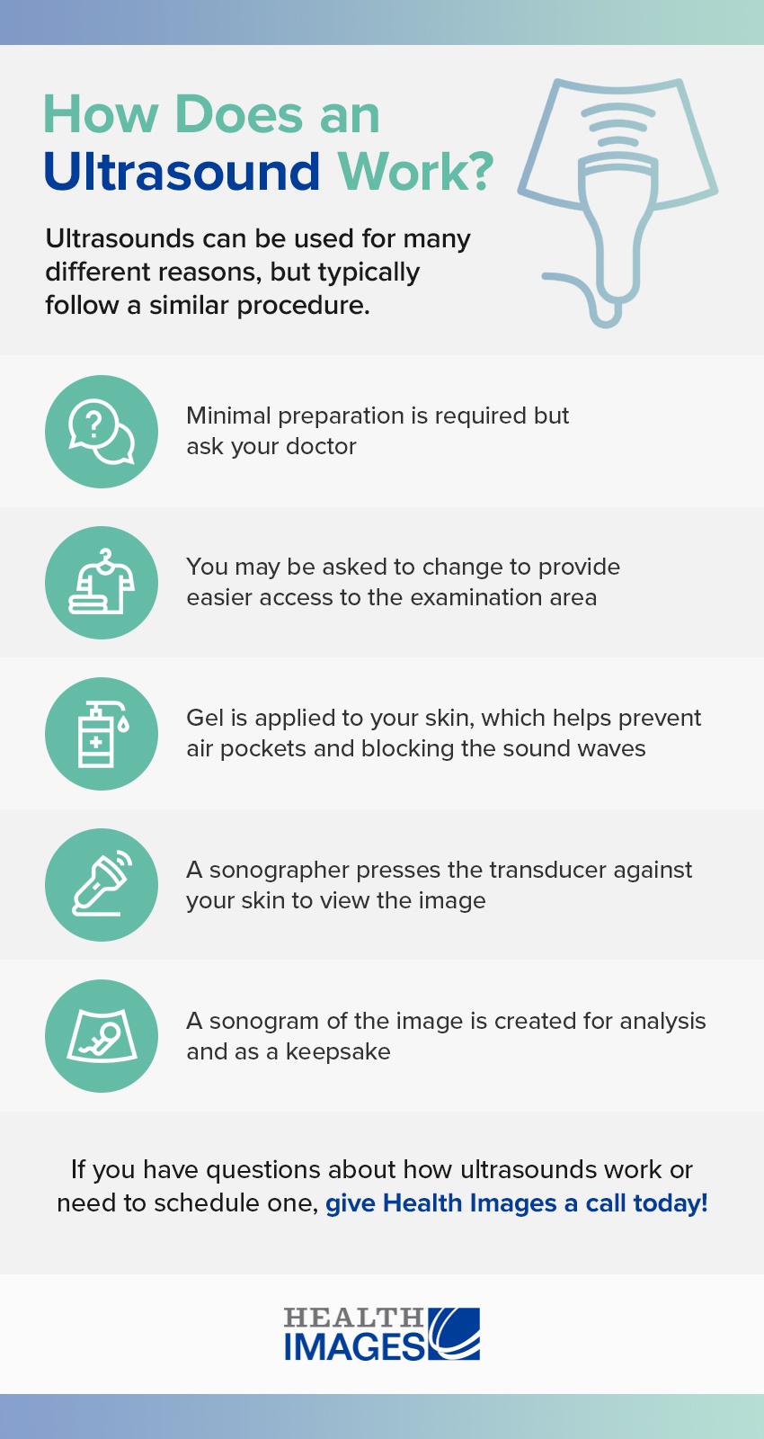
A medical ultrasound is a type of imaging test. Unlike many other imaging tests, ultrasounds do not use radiation to produce images of the internal body. Instead, ultrasounds use high-frequency sound waves to create pictures. An ultrasound is a safe, painless, noninvasive procedure that’s been used in medicine since the 1950s. An ultrasound machine includes a computer, monitor, printer and handheld transducer that sends and receives sound waves.How Does an Ultrasound Work?
In general, an ultrasound machine measures sound waves that have bounced off the body’s internal tissues. The ultrasound’s computer processes these sound waves and turns them into real-time images. If you need to get an ultrasound, you can expect the following steps:- Gel application: Before an ultrasound, the technologist will apply a special gel to the area being studied. The gel is an important part of the ultrasound procedure because sound waves do not travel well through the air. The gel helps remove air and connect the transducer to the skin. As a result, sound waves can travel directly to and from the transducer.
- Use of the transducer wand: The technologist will press the transducer against the gel-covered region of the patient’s skin. The transducer sends high-frequency sound waves into the body. As the sound waves bounce off of organs, tissues and fluids, the transducer picks up subtle changes in the pitch and direction of the sound waves. This information is measured instantly and displayed on the computer.
- Viewing the sonogram: The technologist will save or print the images for interpretation. A radiologist, who is a doctor trained to analyze imaging results, will interpret the sonogram. They will then share their findings with the doctor who requested the exam.
What Is an Ultrasound Used For?
An ultrasound has many uses in the medical world. Doctors may order an ultrasound to check for swollen, damaged or infected organs if a patient is experiencing pain or other symptoms. An ultrasound is a safe, quick way for doctors to check the condition of many organs in the body, such as the:- Heart
- Liver
- Bladder
- Spleen
- Gallbladder
- Uterus
- Ovaries
- Thyroid
- Pancreas
- Kidneys
- Blood clots
- Reduced or increased blood flow to organs
- Tumors
What’s the Difference Between a Sonogram and an Ultrasound?
The easiest way to remember the difference between a sonogram and an ultrasound is to keep in mind that the ultrasound is the procedure, and the sonogram is the result. One could not exist without the other.How Are Sonograms and Ultrasounds Similar?
The sonogram and the ultrasound machine are both components of a highly useful medical exam. As a whole, an ultrasound and the sonogram it produces are beneficial to patients because they are:- Painless
- Radiation-free
- Affordable
- Widely available
- Versatile
- Noninvasive

Contact Health Images Today
Now that you know the difference between a sonogram and an ultrasound, are you ready for the next step? If you need an ultrasound, we’re here to help. At Health Images, we offer ultrasounds and other medical imaging services using the latest technology. Our team of trained technologists will strive to make you feel comfortable and cared for throughout the entire process, and we will work with your schedule to make the experience as quick and convenient as possible. To learn more about our services or to schedule an appointment, contact us today.Call To Schedule Your Appointment
Sources:- https://americanpregnancy.org/prenatal-testing/ultrasound/
- https://www.healthline.com/health/sonogram-vs-ultrasound#how-ultrasound-works
- https://www.radiologyinfo.org/en/info.cfm?pg=genus
- https://www.ncbi.nlm.nih.gov/pmc/articles/PMC3987368/
- https://www.thebump.com/a/sonogram-vs-ultrasound
- https://www.mayoclinic.org/tests-procedures/fetal-ultrasound/about/pac-20394149
- https://www.livestrong.com/article/277225-how-to-read-a-baby-sonogram/
- http://www.meddean.luc.edu/lumen/MedEd/urology/ushome.htm
- https://www.healthimages.com/about-us/
- https://www.whattoexpect.com/pregnancy/pregnancy-health/prenatal-testing-ultrasound/
- https://www.healthimages.com/services/ultrasound-sonogram/
- https://www.ultrasoundtechniciancenter.org/ultrasound-knowledge/how-ultrasound-machines-work.html
- https://blog.cincinnatichildrens.org/radiology/ultrasound-gel-a-necessary-mess
- https://www.healthimages.com/locations/